


Cardiopulmonary exercise testing has long been a cardiologist’s tool. Today, it’s becoming the gold standard for understanding, protecting, and unlocking human athletes.
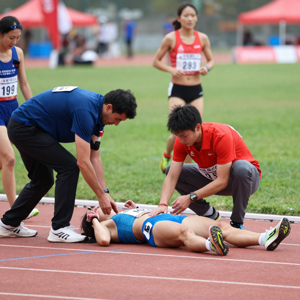
When a 22-year-old long-distance runner collapsed mid-race in a regional championship last season, every standard cardiac screen came back normal. ECG — clean. Echocardiogram — unremarkable. Resting blood work — within range. Yet something was clearly wrong. It wasn’t until her physician ordered a cardiopulmonary exercise test that the truth emerged: a severe ventilatory inefficiency pattern that no resting investigation could have detected.
Stories like hers are becoming more common in sports medicine clinics worldwide — and they’re accelerating a long-overdue shift in how we think about athlete assessment. Cardiopulmonary exercise testing (CPET) is no longer just a tool for cardiac rehabilitation wards. It has arrived, decisively, at the intersection of elite performance and clinical sport science.
“CPET doesn’t just tell you what an athlete can do. It tells you exactly where, and why, their physiology breaks down under stress.”
— Sports cardiology consensus statement, 2024
What CPET actually measures
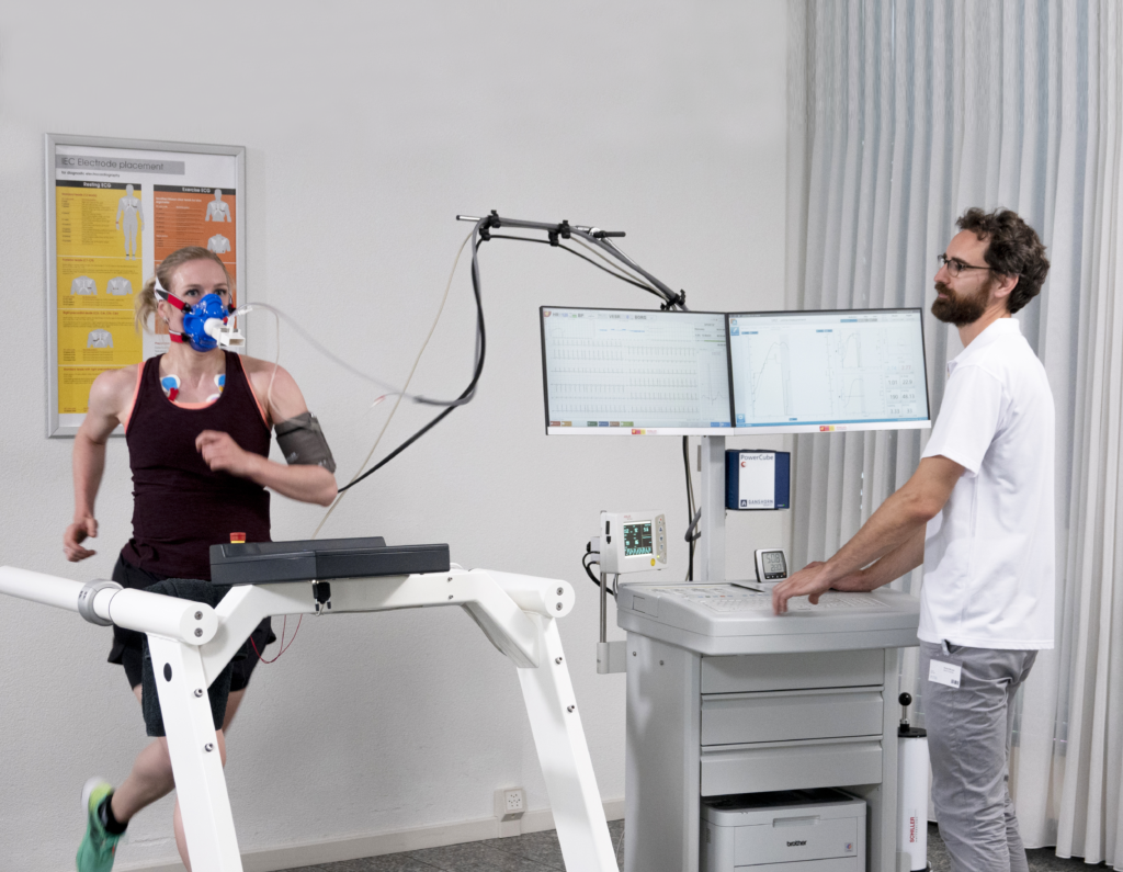
At its core, CPET is an incremental exercise test — typically performed on a treadmill or cycle ergometer — during which the patient breathes through a metabolic analyzer. Unlike a standard stress ECG, which monitors heart rate and electrical activity during exertion but stops there, CPET layers the full respiratory and metabolic response on top: breath-by-breath gas exchange, ventilatory efficiency, and oxygen kinetics — all captured simultaneously in real time. The result is a window into the integrated cardiovascular, pulmonary, and muscular system that no single-system test can replicate.
Key parameters measured during CPET | |
VO₂ max | Peak oxygen consumption; the ceiling of aerobic capacity |
Anaerobic threshold (AT) | The point where lactate begins to accumulate faster than clearance |
VE/VCO₂ slope | Ventilatory efficiency; a sensitive marker of cardiac and pulmonary stress |
Oxygen pulse (VO₂/HR) | Surrogate for stroke volume and cardiac output |
Breathing reserve | Distinguishes exercise limitation as cardiac vs pulmonary in origin |
SpO₂ during exertion | Detects exercise-induced arterial hypoxaemia |
This multi-system view is what gives CPET its diagnostic power. A stress ECG can detect ischaemic changes and arrhythmias provoked by exertion — valuable, but binary. CPET reveals not just whether the system fails, but which component fails, at what intensity, and by how much. It’s the difference between a warning light on a dashboard and a full engine diagnostic readout.
The performance application: precision training zones
For clinicians working with competitive athletes, one of the most practical applications of CPET is the accurate identification of training thresholds. The anaerobic threshold — or more precisely, the ventilatory threshold (VT1 and VT2) — is the cornerstone of evidence-based periodisation.
Traditional field tests, like lactate step testing or time trials, offer useful approximations. But they are prone to hydration status, motivation, and pacing errors. CPET-derived thresholds are physiologically anchored, reproducible, and independent of athlete effort strategy. Coaches and performance scientists who receive CPET data can prescribe training intensities with a granularity that simply wasn’t possible before.
~15% Improvement in training efficiency when zones are CPET-derived vs estimated
3 in 10 Elite athletes show clinically relevant findings only detectable during maximal exertion
40+ Distinct physiological variables captured in a single CPET session
Safety screening: the silent threat problem
Sudden cardiac death in young athletes, though rare, remains one of the most devastating outcomes in sports medicine. Most pre-participation screening protocols rely on history, physical examination, and in many countries, a resting ECG. These are valuable but incomplete.
Conditions such as arrhythmogenic right ventricular cardiomyopathy (ARVC), hypertrophic cardiomyopathy (HCM) with dynamic outflow obstruction, and anomalous coronary artery origins can produce unremarkable stress ECG findings even at near-maximal intensities. CPET, by simultaneously interrogating respiratory and metabolic responses alongside cardiac electrical activity, can unmask ventilatory patterns, abnormal blood pressure trajectories, and oxygen extraction impairments that a stress ECG simply has no mechanism to detect.
In athletes with known structural heart disease, CPET is increasingly used not only for clearance decisions but for ongoing exercise prescription — defining the upper boundary of safe training intensity on an individual, quantified basis rather than empirical guess.
“The question is no longer whether the athlete has a condition. It is whether they can exercise safely, and at what intensity. CPET answers both.”
Return to sport after illness or injury
The post-COVID era has placed CPET in the spotlight in an entirely new context. Many athletes recovering from long COVID present with exercise intolerance, breathlessness, and profound fatigue — yet stress ECGs and standard investigations remain normal. CPET has proven uniquely capable of characterising the physiological patterns underpinning this syndrome: chronotropic incompetence, excessive ventilatory response, and impaired oxygen extraction, among others.
Beyond COVID, CPET is increasingly integrated into return-to-sport protocols following cardiac events, prolonged illness, or major surgery. Objective, quantified exercise tolerance data allows medical teams to make clearance decisions that are defensible, individualized, and safe — moving away from the ambiguous “symptom-free for two weeks” benchmarks that have historically governed these decisions.
Challenges and the road ahead
Despite its growing application, CPET remains underutilised in sports medicine — largely due to equipment cost, the need for trained interpreters, and a lack of standardised protocols adapted specifically for athletic populations. Most reference ranges were developed in sedentary or clinical cohorts, and their direct application to trained athletes requires careful qualification.
There is also the question of access. At present, CPET is predominantly available in tertiary sports medicine centres, university research labs, and elite sporting organisations. Expanding availability to community and amateur athletes — who arguably carry a higher undetected risk burden — remains an important public health challenge.
Work is ongoing to develop sport-specific normative data, simplified reporting frameworks for non-specialist clinicians, and portable CPET systems that can be deployed in field settings. The trajectory is clearly toward broader adoption; the question is how quickly the infrastructure can catch up.
For the runner who collapsed at that regional championship, a CPET didn’t just provide a diagnosis. It provided a roadmap — a precise, quantified understanding of where her physiology was failing and at what intensity she could safely resume training. She competed again six months later.
That is what CPET offers sports medicine: not just a test, but a conversation between the clinician, the data, and the athlete — conducted at the only moment that truly matters: when the body is fully asked to perform.